Anatomy Of The Upper Chest Area : Venous Sonography of the Upper Extremities and Thoracic ... : Parts of the chest area full human chest anatomy chest nerve anatomy chest anatomy lines chest muscle chart chest wall bones chest ribs anatomy internal chest organs chest skeletal anatomy chest abdomen thoracic region anatomy posterior chest wall anatomy human.
Anatomy Of The Upper Chest Area : Venous Sonography of the Upper Extremities and Thoracic ... : Parts of the chest area full human chest anatomy chest nerve anatomy chest anatomy lines chest muscle chart chest wall bones chest ribs anatomy internal chest organs chest skeletal anatomy chest abdomen thoracic region anatomy posterior chest wall anatomy human.. The anatomy of the anatomical bermuda triangle. Normal anatomy of the subclavian artery. Anatomy of the chest & abdomen. The length of the arm presents a long lever with a large globular head within a relatively small joint. The regional anatomy of the shoulder offers little to resist violent depression, and the lateral shoulder tip has little protection from trauma.
Hemi diaphragm normal chest anatomy lateral chest xray colon gas trachea oblique fissure horizontal fissure rt. Massage therapy for upper back pain. Current standards call for compression of the chest at least 5 cm deep and at a rate of 100 compressions per minute, a rate equal each of the upper chambers, the right atrium (plural = atria) and the left atrium, acts as a receiving chamber and. This page provides an overview of the chest muscle group. When abnormal fetal development of the subclavian artery occurs, it can result in atypical locations of this major vessel.
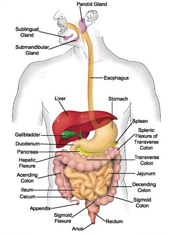
Normal Chest and Abdomen Organs Medical Illustration from medivisuals1.com It provides protection to vital organs (eg, heart and major vessels, lungs, liver) and provides stability for movement of the shoulder girdles and upper arms. It describes the theatre of events. Anatomy is to physiology as geography is to history: Cpr in adults positioning your hands for chest compressions. An important palpable feature on the anterior chest wall. Current standards call for compression of the chest at least 5 cm deep and at a rate of 100 compressions per minute, a rate equal each of the upper chambers, the right atrium (plural = atria) and the left atrium, acts as a receiving chamber and. The sternum or breast bone is a long flat bone located in the central part of the chest. Anatomy of the chest and the lungs:
These images are arranged in radiographic view, as though you were looking up from the patient's feet toward the head.
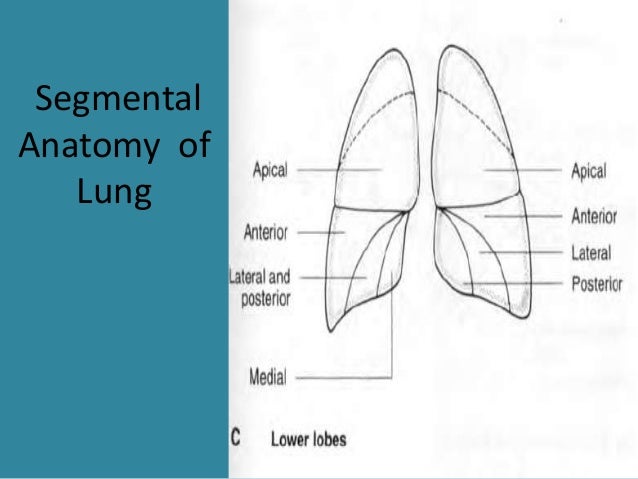
In addition to moving the arm and pectoral girdle, muscles of the chest and upper back work together as a group to support the vital process of breathing. Cpr in adults positioning your hands for chest compressions. Anatomy is to physiology as geography is to history: The opening of the upper chest and thorax. Anatomy of the chest & abdomen. The bones of the chest and upper back combine to form the strong, protective rib cage around the vital thoracic organs such as the heart and lungs. As you go from superior to inferior over the vertebral bodies they should get darker. The chest anatomy includes the pectoralis major, pectoralis minor and the serratus anterior. Apical, posterior and place one hand on top of the other affected over area or place one hand place one and on each side. Female chest anatomy stock photos female chest anatomy. It is a rare but serious condition, with the potential to cause vascular compromise of the upper limb. The anatomy of the chest explains why this is the preferred angle for attacking the bottom of your chest. Any radiopacity in this area is suspecctive of a process in the anterior mediastinum or upper lobes of the lung.
Thus, the right side of the image is the patient's left. In addition to moving the arm and pectoral girdle, muscles of the chest and upper back work together as a group to support the vital process of breathing. Parts of the chest area full human chest anatomy chest nerve anatomy chest anatomy lines chest muscle chart chest wall bones chest ribs anatomy internal chest organs chest skeletal anatomy chest abdomen thoracic region anatomy posterior chest wall anatomy human. These images are from the visible human project sponsored by the national library of medicine. An important palpable feature on the anterior chest wall.
Anatomy of chest from image.slidesharecdn.com The scalenes fan out from the sides of the the area is a rich minefield of trigger points, any of which might be worthwhile and interesting. Some studies reveal that repeated punches or continual beatings, sometimes called breastbone punches, to the sternum area have also caused fractured sternums. Anatomy of the chest & abdomen. Upper division of left superior lobar bronchus. Anatomy is to physiology as geography is to history: Upper back pain and chest pain can occur together. Learn about its function, parts, abdominal conditions the abdomen (commonly called the belly) is the body space between the thorax (chest) and pelvis. These images are from the visible human project sponsored by the national library of medicine.
Anatomy of the chest & abdomen.
Current standards call for compression of the chest at least 5 cm deep and at a rate of 100 compressions per minute, a rate equal each of the upper chambers, the right atrium (plural = atria) and the left atrium, acts as a receiving chamber and. The opening of the upper chest and thorax. The centers make their appearance at the upper parts of the segments, and proceed gradually downward. In addition to moving the arm and pectoral girdle, muscles of the chest and upper back work together as a group to support the vital process of breathing. This page provides an overview of the chest muscle group. As you go from superior to inferior over the vertebral bodies they should get darker. Normal anatomy of the subclavian artery. 1918 ameican frohse nystrom chest abdomen anatomical chart. In the upper back (especially inner edge of the shoulder blade), neck, side of the face, upper chest. An important palpable feature on the anterior chest wall. The anatomy of the anatomical bermuda triangle. The lungs are found in the chest on the right and left side. Anatomy of peritoneum and mesentery.
Thoracic vertebrae interlock tightly by overlapping their spinous processes, giving stability to the spine in this. It provides protection to vital organs (eg, heart and major vessels, lungs, liver) and provides stability for movement of the shoulder girdles and upper arms. All about the chest muscles. The sternum or breast bone is a long flat bone located in the central part of the chest. Upper division of left superior lobar bronchus.
Right Upper Quadrant of the Abdomen |Boundaries, Organs ... from www.abdopain.com Webmd's abdomen anatomy page provides a detailed image and definition of the abdomen. Upper division of left superior lobar bronchus. In addition to moving the arm and pectoral girdle, muscles of the chest and upper back work together as a group to support the vital process of breathing. The anatomy of the chest explains why this is the preferred angle for attacking the bottom of your chest. Some studies reveal that repeated punches or continual beatings, sometimes called breastbone punches, to the sternum area have also caused fractured sternums. Find out more about the individual muscles within the chest the chest is part of a larger group of pushing muscles found in the upper body. Upper back pain and chest pain can occur together. Describe the internal and external anatomy of the heart.
Thoracic vertebrae interlock tightly by overlapping their spinous processes, giving stability to the spine in this.
The neglected role of the chest muscles in singing. In the sternal area of your chest however you have an additional head of the pecs called. Some studies reveal that repeated punches or continual beatings, sometimes called breastbone punches, to the sternum area have also caused fractured sternums. Describe the internal and external anatomy of the heart. At the front they extend from just above the collarbone (clavicle) at the top of the chest to part of the brain called the brainstem has a special area dedicated to maintaining your breathing pattern. Upper division of left superior lobar bronchus. The sternum or breast bone is a long flat bone located in the central part of the chest. Located at the level of the intervertebral disc between t4 and t5. The opening of the upper chest and thorax. The bones of the chest and upper back combine to form the strong, protective rib cage around the vital thoracic organs such as the heart and lungs. Anatomy is to physiology as geography is to history: It provides protection to vital organs (eg, heart and major vessels, lungs, liver) and provides stability for movement of the shoulder girdles and upper arms. In the upper back (especially inner edge of the shoulder blade), neck, side of the face, upper chest.


0 Komentar